View a slide
Clicking an image file in the object browser opens the image viewer, where you can examine whole-slide scans at any zoom level.
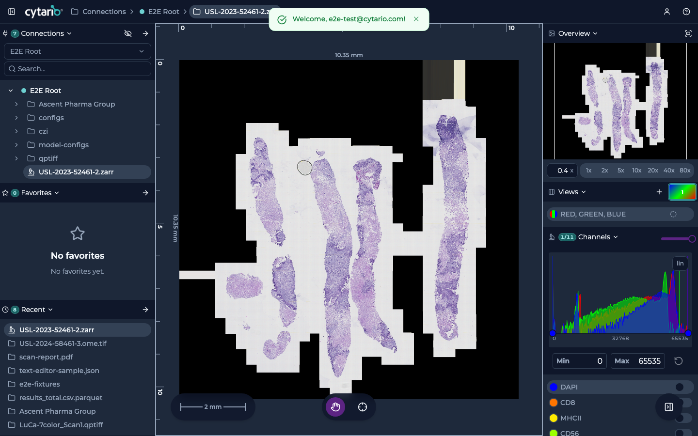
Moving around the image
- Pan by dragging the image.
- Zoom with the scroll wheel or the magnification presets (for example 5×, 10×, 20×) at the top of the viewer.
- The navigator (miniature map) shows where you are within the whole slide; click or drag it to jump around.
- Use Reset View State to return to the starting view.
Channels and colors
- For multi-channel fluorescence (mIF) images, the viewer shows the channels as tabs or presets; select them to compose the combination you want to examine.
- Adjust per-channel contrast and brightness to optimize signal visibility, and apply pseudo-colors to distinguish overlapping markers.
- The Channels / Overlays / Annotations panel toggles each layer and its opacity.
What's next
- Analyze cell-segmentation overlays and query tabular data on the same image.
- Run analysis on an image or folder if your organization has compute enabled.
These deeper workflows are covered in the screens and reference sections of this guide.