Viewing slides
Clicking an image in the object browser opens it in the image viewer. The viewer renders whole-slide scans at full resolution, with pan, zoom, and magnification controls, and supports both brightfield (H&E / IHC) and multi-channel fluorescence (mIF) images.
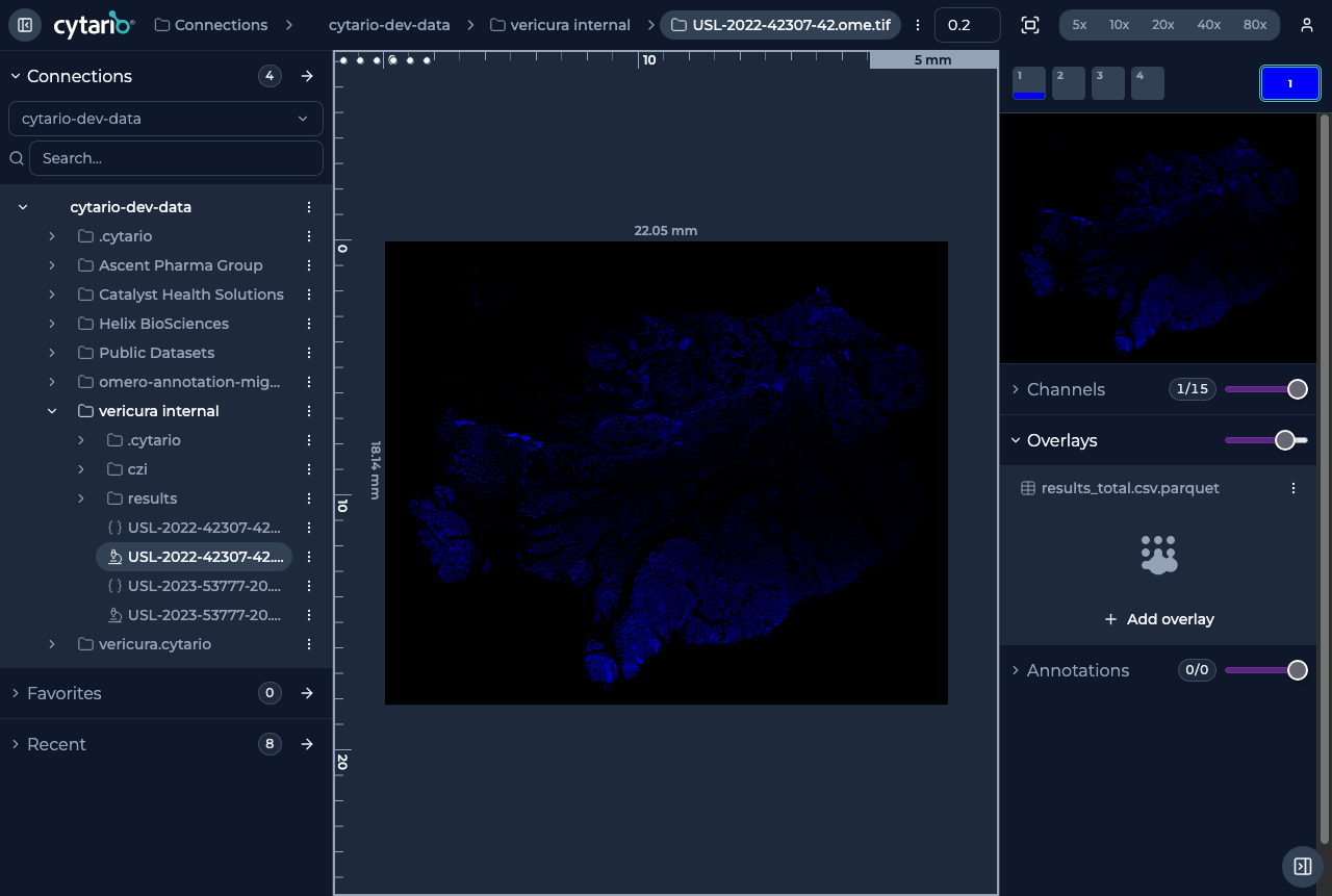
Moving around
- Pan by dragging the image.
- Zoom with the scroll wheel or the magnification presets at the top of the viewer (5x / 10x / 20x / 40x / 80x).
- A navigator (miniature map) shows where you are within the whole slide; click or drag it to jump to another region.
- Reset View State returns the view to the default position and zoom.
Measuring and orienting
The viewer shows physical-dimension information (a scale) so you can gauge feature sizes while you inspect tissue morphology. Spatial measurements and side-by-side comparison are part of routine viewing.
- The scale bar and rulers are labelled in metric units (mm / µm) by default. Click the scale bar to switch the whole viewer between metric units and image pixels — the choice is remembered across sessions.
Image controls
An Image controls panel to the side of the viewer lets you manage the layers of the image:
| Layer | What it controls |
|---|---|
| Channels | Visibility and opacity of image channels |
| Overlays | Cell-segmentation overlays and their fill opacity |
| Annotations | Drawn annotations and their opacity |
Each layer can be toggled on or off, so you can inspect the raw image, its segmentation overlay, or both together.
A Settings section at the bottom of the panel collects viewer display options: show or hide the scale bar and the rulers, and switch the display unit between metric and pixels. The unit choice is the same toggle as clicking the scale bar and is remembered across sessions.
In this section
- Channels & colors — composing channels and adjusting contrast, brightness, and pseudo-color.
- Overlays & quantitative analysis — loading cell-segmentation data and querying it.
- Annotations — drawing on images.